The Shadow and the Light: Rosalind Franklin, Photo 51, and the Unveiling of Life\'s Blueprint
A meticulously crafted X-ray diffraction image, born from a humid London laboratory in May of 1952, holds within its crystalline patterns the very essence of life. This unassuming photograph, designated \"Photo 51,\" was the culmination of years of painstaking scientific endeavor by Dr. Rosalind Franklin, a brilliant and often underestimated crystallographer. It was this image, more than any other single piece of evidence, that illuminated the fundamental architecture of deoxyribonucleic acid (DNA) and provided the critical, irrefutable clues that would ultimately lead to the groundbreaking discovery of the double helix by James Watson and Francis Crick. Yet, the story of Photo 51 and its rightful place in scientific history is one intertwined with intellectual property, professional rivalries, and a lingering injustice that continues to be re-examined decades later.
The mid-20th century was a fertile ground for scientific exploration. The post-war era saw an explosion of research across numerous fields, driven by both a thirst for fundamental knowledge and the pragmatic applications that such knowledge could unlock. In biology, the question of heredity, the mechanism by which traits are passed from parents to offspring, loomed large. While Gregor Mendel had laid the groundwork in the 19th century with his laws of inheritance, the molecular basis of these laws remained elusive. Scientists hypothesized that a complex molecule, likely found within the nucleus of every cell, was responsible for carrying this genetic information. Proteins, with their diverse amino acid sequences, were initially considered strong contenders, but as research progressed, deoxyribonucleic acid (DNA), a simpler molecule composed of nucleotides, began to emerge as a more plausible candidate.
The chemical composition of DNA was largely understood by the 1940s. It was known to be a polymer made up of repeating units called nucleotides, each consisting of a phosphate group, a deoxyribose sugar, and one of four nitrogenous bases: adenine (A), guanine (G), cytosine (C), and thymine (T). The arrangement of these bases, it was theorized, must hold the key to the genetic code. However, understanding the *structure* of DNA was paramount to understanding its *function*. How did this linear sequence of bases translate into the complex instructions that govern all living organisms?
Enter Rosalind Franklin. Born in London in 1920, Franklin displayed an extraordinary aptitude for science from a young age. She attended St Paul\'s Girls\' School, one of the few schools to teach physics and chemistry to girls, where she excelled. Her academic brilliance continued at Newnham College, Cambridge, where she studied natural sciences. While her contemporaries, like Watson and Crick, were immersed in the theoretical aspects of molecular biology at Cambridge, Franklin pursued a more experimentally grounded approach. She earned her PhD in physical chemistry from Cambridge in 1945, with her research focusing on the porosity of coal, a vital subject for the war effort. This early work honed her skills in X-ray diffraction, a powerful technique that uses the scattering of X-rays by atoms in a crystal to deduce the arrangement of those atoms – essentially, to determine molecular structure.
After her postdoctoral work in Paris, where she further refined her X-ray crystallography techniques, Franklin was recruited to King\'s College London in 1951 to work in the Medical Research Council (MRC) Unit for Molecular Biology. Her mission was to apply her expertise to the study of DNA. This was a pivotal moment, both for Franklin and for the field of molecular biology. She was tasked with obtaining high-resolution X-ray diffraction images of DNA fibers, a notoriously difficult undertaking. DNA molecules are long and thin, and coaxing them into ordered, crystalline fibers that could be bombarded with X-rays required immense skill and patience.
Franklin’s laboratory at King’s College was a crucial environment. She was joined by a young research assistant, Raymond Gosling, who became instrumental in her work. Together, they embarked on the meticulous process of preparing DNA samples and exposing them to X-ray beams. The technique involves mounting a thin fiber of DNA, often prepared by stretching it under tension, in a small, airtight chamber. An X-ray beam is then directed at the fiber, and the diffracted X-rays are recorded on photographic film. The pattern of spots and streaks on the film, known as a diffraction pattern, is a unique fingerprint of the molecule’s three-dimensional structure.
Franklin’s approach was characterized by her rigorous scientific methodology and her insistence on precise, quantitative data. Unlike some of her contemporaries who favored speculative model-building, Franklin believed that empirical evidence should guide theoretical interpretation. She was also meticulous in her preparation of the DNA fibers. She realized that the hydration level of the DNA was critical and that under different humidity conditions, DNA fibers could adopt distinct forms. She identified two main forms, which she labeled \"A\" and \"B.\" The \"A\" form, obtained under drier conditions, showed a different diffraction pattern, while the \"B\" form, obtained under more humid conditions, was more characteristic of DNA in a hydrated biological environment. It was the \"B\" form that held the key to DNA’s biological structure.
The work in Franklin’s lab was demanding. The X-ray diffraction process was slow, requiring long exposure times, sometimes days. The data generated was complex, and its interpretation demanded a deep understanding of diffraction theory and mathematical analysis. Franklin was a solitary figure in many ways, not easily swayed by the prevailing scientific winds or the social dynamics of the lab. Her directness and no-nonsense approach sometimes clashed with the more collegial atmosphere of other research groups.
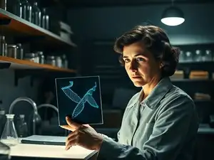
In May 1952, after countless hours of refinement and experimentation, Franklin and Gosling obtained a particularly clear and striking diffraction pattern from a well-oriented DNA fiber of the \"B\" form. This was Photo 51. The image displayed a distinct ‘X’ shape, a hallmark of helical structures, with a series of precise spots arranged around its perimeter. The central cross was unmistakable, strongly suggesting a helical molecule, and the spacing of the spots provided crucial information about the dimensions of this helix. The presence of the \"meridian\" spots indicated the helical repeat, while the \"equator\" spots provided information about the base pairing and the overall diameter of the molecule.
Franklin’s interpretation of Photo 51 was thorough and insightful. She concluded that DNA was a helix, with the phosphate backbone located on the outside of the molecule, a critical deduction. She also meticulously calculated the precise dimensions of the helix: its diameter, the distance between the bases along the axis, and the pitch of the helix (the distance for one complete turn). Her unpublished reports and lab notebooks contain detailed analyses and measurements that were remarkably accurate. She estimated the B-DNA helix to be approximately 2 nanometers in diameter, with a helical repeat of 3.4 nanometers, and about 10 base pairs per turn. These were not guesses; they were the result of rigorous mathematical analysis of the diffraction data.
However, the story of Photo 51 is inextricably linked to the events that unfolded at the Cavendish Laboratory in Cambridge, where James Watson and Francis Crick were also attempting to build a model of DNA. Watson, a young American biologist, and Crick, a British physicist, were working on a more theoretical, model-building approach, heavily influenced by Linus Pauling’s work on protein structures. Their early models were flawed, reflecting a lack of experimental data.
The transfer of crucial information from Franklin’s lab to Watson and Crick remains a contentious point in the history of science. Maurice Wilkins, a colleague of Franklin’s at King\'s College and a rival for control of the DNA research program, had shown Photo 51 to Watson without Franklin’s explicit knowledge or consent. Wilkins was himself using X-ray diffraction to study DNA, and his research was funded by the same MRC unit as Franklin’s. While there might have been an understanding that their research was collaborative, the unauthorized sharing of such a pivotal piece of evidence is ethically questionable.
Furthermore, Max Perutz, the chairman of the MRC unit and a Nobel laureate, had access to Franklin’s detailed reports, which included her quantitative analyses of Photo 51. These reports were circulated among researchers, including Watson and Crick, as part of the unit\'s internal communication. Franklin, being somewhat of an outsider in the predominantly male scientific establishment and perhaps not as adept at navigating academic politics, did not aggressively pursue the publication of her findings before her untimely death.
Watson and Crick, armed with Photo 51 and Franklin\'s crucial quantitative data (which they later admitted was instrumental), were able to refine their model building. The “X” shape of Photo 51 strongly suggested a helical structure. Franklin’s measurements confirmed the dimensions of this helix and, critically, indicated that the phosphate backbone was on the outside. This placement of the backbone was a vital constraint for their model. Coupled with Erwin Chargaff\'s rules, which stated that in DNA, the amount of adenine (A) always equals the amount of thymine (T), and the amount of guanine (G) always equals the amount of cytosine (C), Watson and Crick were able to conceive of the complementary base pairing (A with T, and G with C). This pairing explained how the genetic information could be copied.
In April 1953, Watson and Crick published their seminal paper in the journal *Nature*, proposing the now-famous double helix model of DNA. The paper was remarkably brief, a mere 900 words, and famously stated, \"It has not escaped our notice that the specific pairing we have postulated immediately suggests a possible copying mechanism for the genetic material.\" This statement, while understated, hinted at the revolutionary implications of their model.
The acknowledgments in the Watson and Crick paper are telling. They thank Maurice Wilkins and his colleagues \"for helpful discussions\" and also acknowledge the \"pre-publication communication of the general nature of the results\" from Franklin and Gosling. This acknowledgment, while present, is widely seen as insufficient given the extent to which Franklin\'s data underpinned their breakthrough. Franklin and Gosling’s own paper, detailing their experimental findings and interpretation of Photo 51, was published in the same issue of *Nature*, but it appeared *after* Watson and Crick\'s model.
The Nobel Prize in Physiology or Medicine in 1962 was awarded to James Watson, Francis Crick, and Maurice Wilkins for their discoveries concerning the molecular structure of nucleic acids and its significance for information transfer in living material. Rosalind Franklin was conspicuously absent from this recognition. Tragically, she had died of ovarian cancer in 1958 at the young age of 37, never knowing the full extent of her contribution or the Nobel Prize awarded to her colleagues.
The story of Rosalind Franklin and Photo 51 is a poignant reminder of the complexities of scientific discovery and the often-unseen contributions of individuals, particularly women, in the scientific arena. Franklin’s meticulous, data-driven approach was the bedrock upon which the double helix model was built. Photo 51 was not merely a pretty picture; it was a Rosetta Stone, a key that unlocked the secret of DNA’s structure.
Franklin’s legacy has been re-evaluated and increasingly recognized over the decades. While Watson and Crick certainly made a brilliant conceptual leap in proposing the double helix, it is undeniable that Franklin’s experimental data provided the crucial scaffolding and validation for their model. Her precise measurements of the B-DNA form, her conclusion that the phosphate backbone was on the exterior, and the very clarity of Photo 51 itself were indispensable.
The impact of Photo 51 cannot be overstated. It provided definitive evidence for the helical nature of DNA, a structure that perfectly explained how genetic information could be stored, copied, and passed down through generations. The double helix model revolutionized biology, opening the door to understanding gene expression, genetic engineering, and a vast array of molecular processes that govern life. It laid the foundation for fields such as genomics, proteomics, and personalized medicine.
Rosalind Franklin\'s personal life was as dedicated to science as her professional life. She was a private individual, passionate about her work, and seemingly less inclined to engage in the social networking and self-promotion that was prevalent in some scientific circles. She was known for her intelligence, her independence, and her unwavering commitment to scientific accuracy. Her early death was a devastating loss to the scientific community.
The narrative surrounding Photo 51 and Franklin’s contribution is a cautionary tale about intellectual property, scientific collaboration, and the biases that can affect the recognition of scientific merit. It highlights the importance of giving credit where credit is due and of acknowledging the foundational experimental work that underpins theoretical breakthroughs.
In recent years, there has been a concerted effort to ensure Rosalind Franklin receives the recognition she deserves. Biographies, documentaries, and academic discussions have brought her story to a wider audience, emphasizing her pivotal role in one of the most significant scientific discoveries of the 20th century. Photo 51, once a somewhat obscure image among a select few, is now recognized as a symbol of Franklin’s genius and her indispensable contribution to our understanding of life itself.
The image itself, a stark and elegant pattern of intersecting lines and dots, continues to fascinate. It is a testament to the power of X-ray crystallography, a technique that allows us to peer into the subatomic world and unravel the intricate architecture of molecules. The ‘X’ at its center is not just a geometric shape; it is a harbinger of life’s most fundamental secret, a blueprint encoded in the very fabric of our being.
Rosalind Franklin’s story serves as a powerful reminder that scientific progress is rarely the work of a single individual. It is often a collaborative effort, built upon the contributions of many. However, it is also a story that underscores the importance of individual brilliance and the often-unheralded efforts of those who lay the groundwork for major discoveries. Photo 51, the image that unlocked DNA, stands as a perpetual monument to Rosalind Franklin’s extraordinary scientific legacy, a legacy that, though delayed, is now firmly etched in the annals of scientific history. The elegant cross, the defining feature of Photo 51, is no longer just a scientific artifact; it is a symbol of truth, perseverance, and the enduring power of meticulous scientific inquiry.